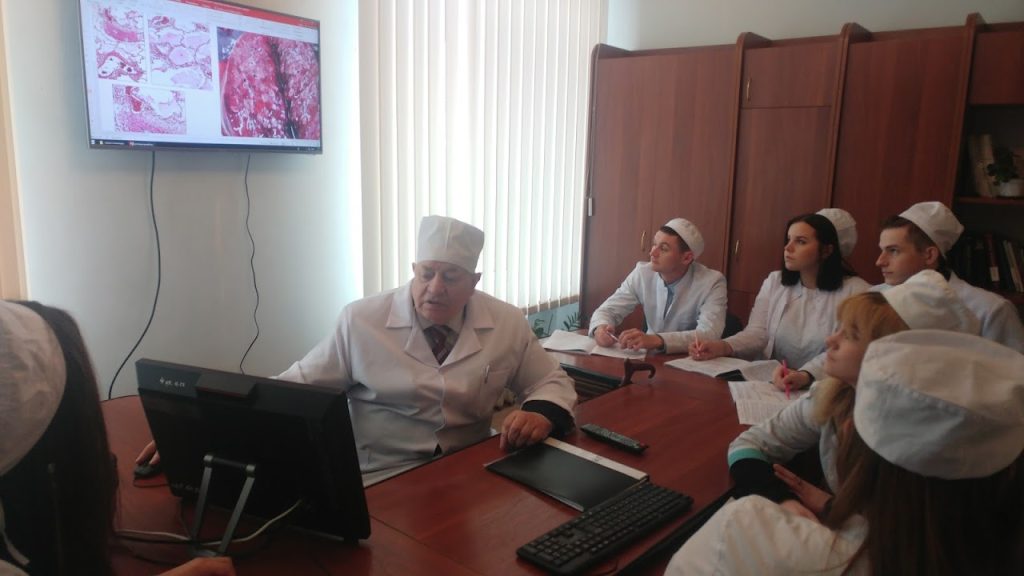
The educational process at the Educational and Scientific Institute of Morphology is organized taking into account the possibilities of modern information technologies of learning and is oriented towards the formation of an educated, harmoniously developed personality, professional mobility and rapid adaptation to changes and development in medicine, in the fields of science, technology and in the social and cultural sphere, in today’s conditions.
All departments of the Educational and Scientific Institute of Morphology train students of higher education in normative and selective academic disciplines. Modern lecture halls are equipped with video systems, mobile interactive whiteboards. Practical classes are held in specially equipped classrooms with the necessary technical teaching aids. Practical classes for students of the 1st – 3rd courses are conducted according to the international system, and for students of the 4th-5th courses, according to the “One Day” method.
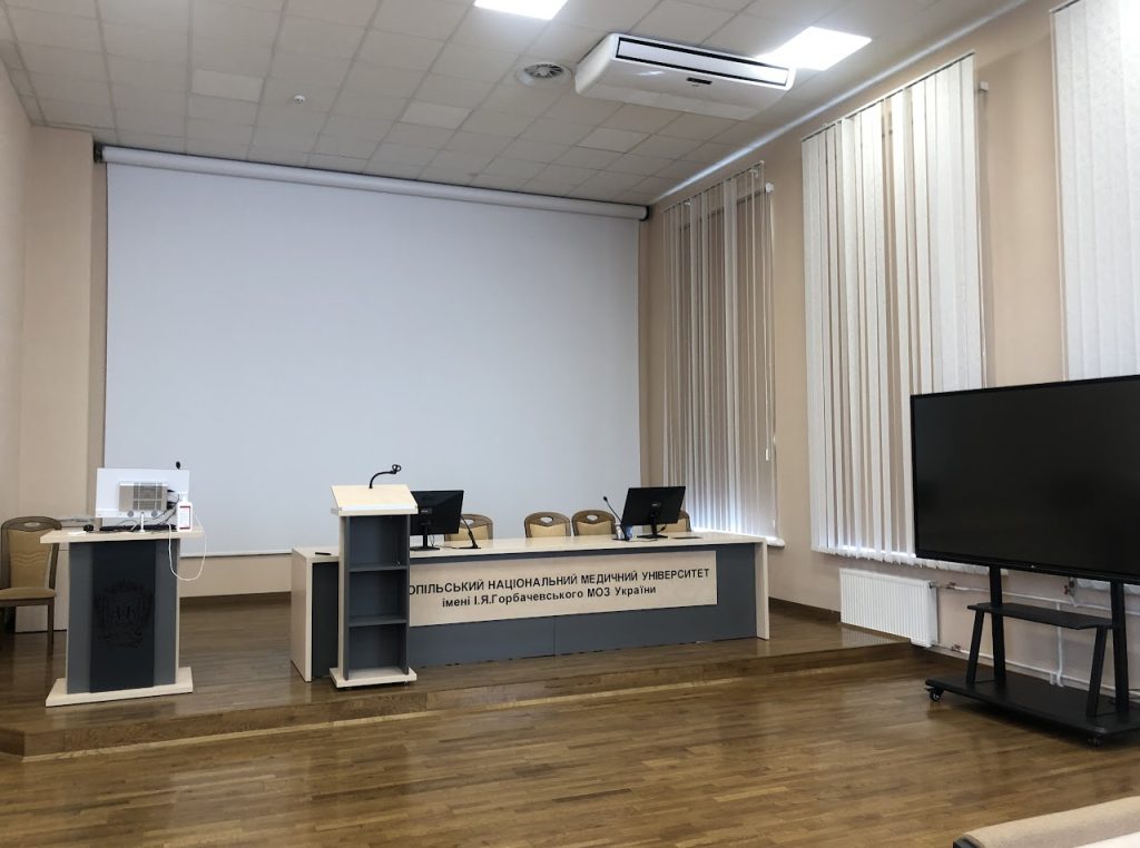
Modern lecture halls waiting for students
A view of the educational facilities of the Institute of Morphology
The Museum of Human Anatomy Department is located on the second floor of the morphological building on the basis of the Department of Human Anatomy, founded in 1959 by Professor Mykola Polyankin. All of the museum’s specimens are rationally systematized in display cabinets according to the functional-anatomical principle and in accordance with curricula and programs on human anatomy. The museum has fully equipped sections with the exposition of educational and scientific specimens and the production of tables on osteoarthrology, splanchnology, angioneurology, peripheral and central nervous systems. The section on teratology “Developmental defects in children” deserves special attention, in which original drugs obtained from clinical institutions of the city of Ternopil are described in details. On 596 specimens (wet and dry), you can see the natural shape of organs, their topographic relationships, projections of blood vessels and nerves, and the structure of individual organs.
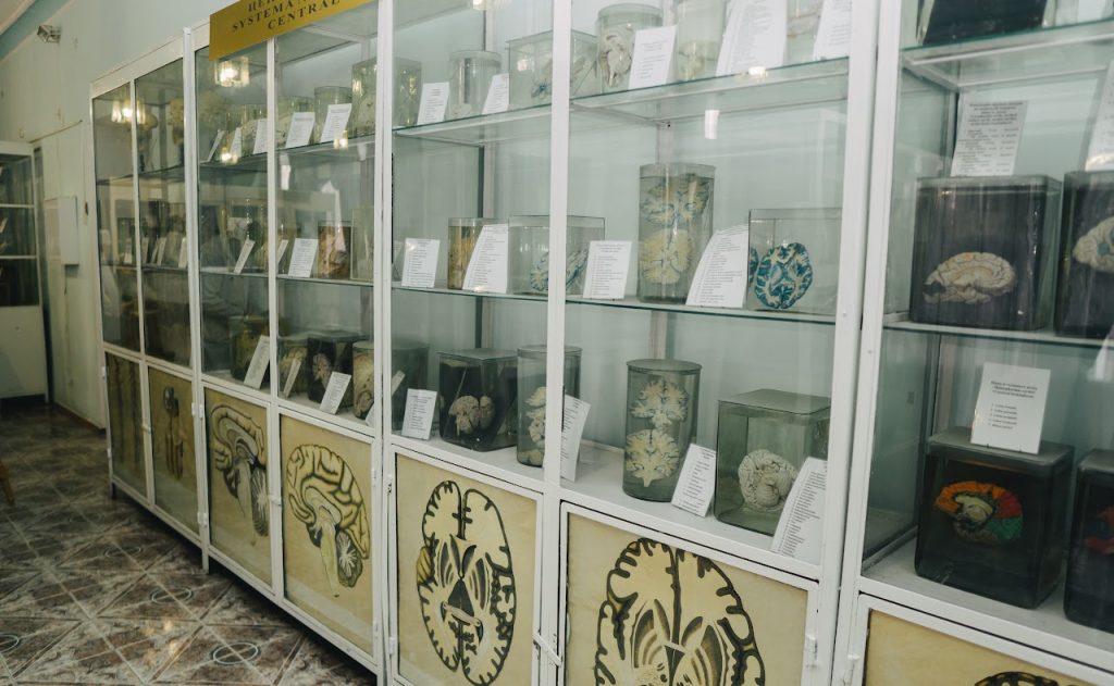
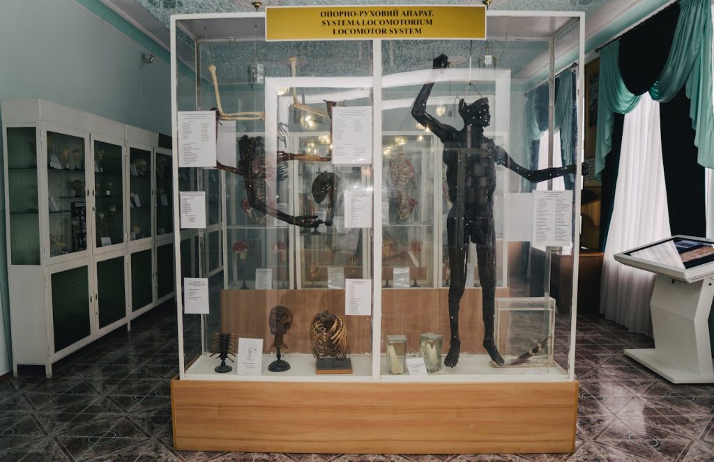
A fragment of the exposition of specimens in the museum of the Human anatomy Department
Museums of the Department of Pathological Anatomy with Autopsy Course and the Department of Forensic Medicine began to be formed from the first day of the foundation of our university, at that time named the Institute. And the immediate founders were associate professor B. Dubchak and assistant S. Abramov, who produced the first hundreds of macrospecimens. Museums of the department are constantly used to form attitudes towards a healthy lifestyle of school and student youth, fight against bad habits, visually reproducing the consequences and complications of alcoholism, drug addiction, smoking, etc. There is also an educational forensic medical museum on the basis of the department, which today has about 1,000 different exhibits from all sections of forensic medicine and is one of the richest specialized medical museums of Ukraine in terms of exposition. In it, along with the expositions of the scene, stands with original samples of various traumatic objects, cold and firearms are displayed.
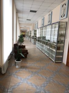
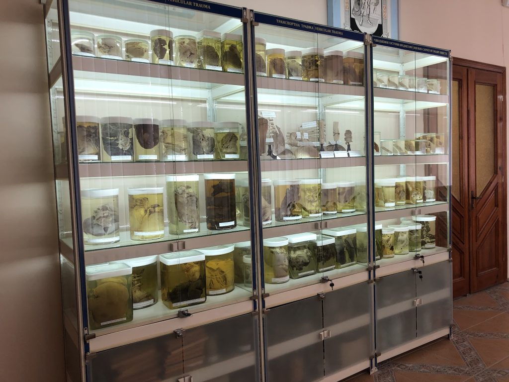
A fragment of the exposition of specimens in the museum of the Pathologic Anatomy with Autopsy course and Forensic Pathology
The Department of Anatomy uses interactive tables (manufactured by Visual fusion technology LLC) with appropriate software, virtual anatomy programs that allow you to view the layer-by-layer structure of the human body, the course of blood vessels, the topography of peripheral nerves, in a three-dimensional 3D mode. bones and their connections. A lot of illustrative material has been integrated into the database: educational videos, atlases and visual video materials on dissection of physical bodies and acquisition of other practical skills. Also, in the database, students can find computer and magnetic resonance tomograms of all normal areas of the human body.
Classes based on the interdepartmental educational and training center with VR software Sharecare YOU, 3D Organon and Cadaver dissection for the comprehensive study of human anatomy have been introduced into the educational process.
The use of the latest digital systems and learning platforms in the educational process at the Department of Human Anatomy
Practical classes at the Department of Histology and Embryology are held in classrooms equipped with video systems, with the help of which teachers demonstrate histological microspecimens. Students study histostructures using modern optical microscopes. An atlas of electron microphotographs is widely used for in-depth study of cells and tissues. The Virtual Microscopy Laboratory (Histology Guide) provides students with a flexible and accessible learning environment.
Typical weekdays at the Department of Histology and Embryology
The main task of the Department of Pathological anatomy with Autopsy Course and Forensic Medicine is to improve the applied study of pathological processes and through clinical and morphological analysis to teach students to evaluate the essence of the disease, its clinical manifestations, as well as to solve the issues of forensic medical examination in cases of violent death. The classrooms of the department are equipped with multimedia equipment and plasma televisions, which provides a demonstration of micropreparations and analysis of pathological changes for the entire audience. The teacher’s workplace is equipped with a computer and a video system, which makes it possible to transfer images from the microscope to the TV screen, to show fragments of video films on the subject of the lesson.
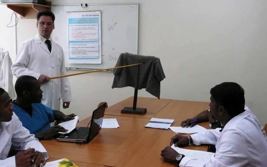
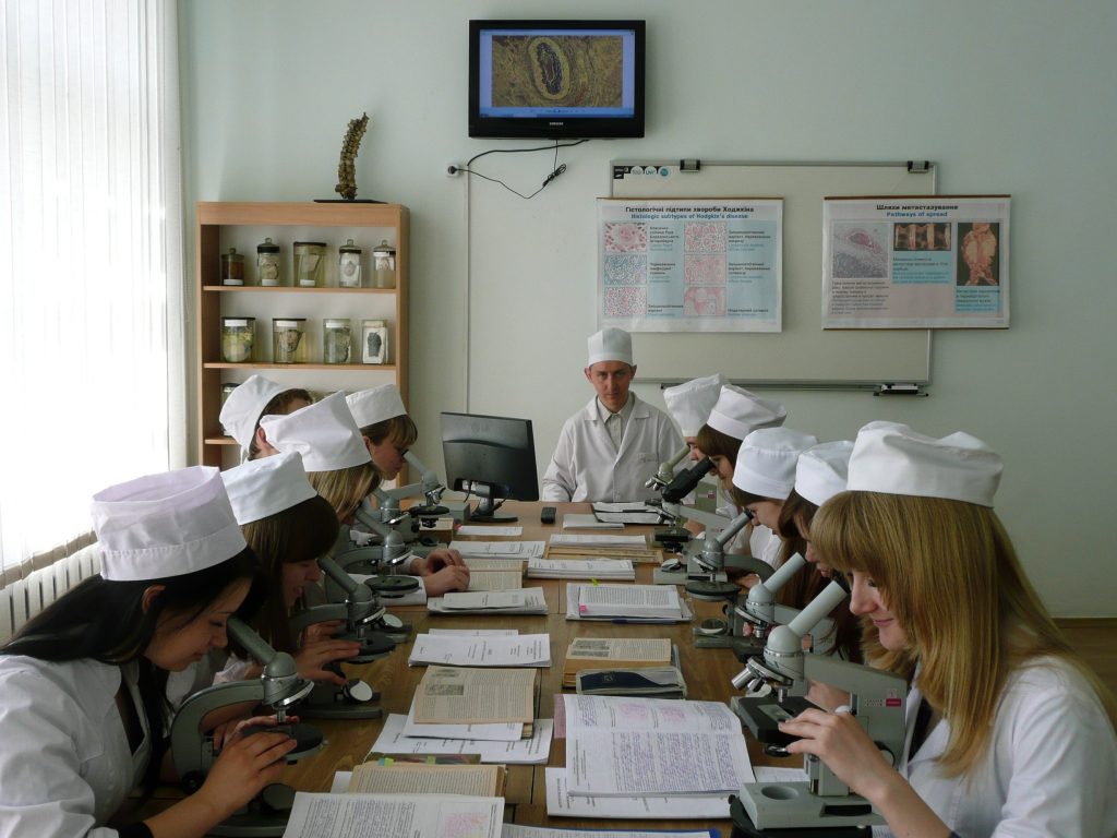
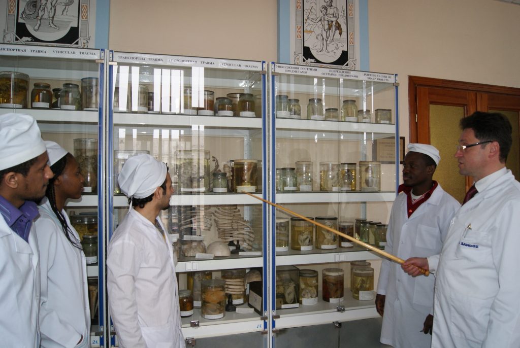
Conducting practical classes by teachers of the Pathologic Anatomy, Autopsy course and Forensic Pathology
Educational process at the department of operative surgery with topographical anatomy
In the conditions of the adaptive quarantine introduced in our country, lectures, practical classes, consultations and exams were held online according to the schedule using Microsoft Teams software.

Online learning process
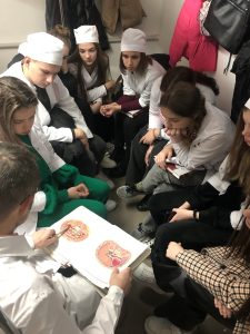
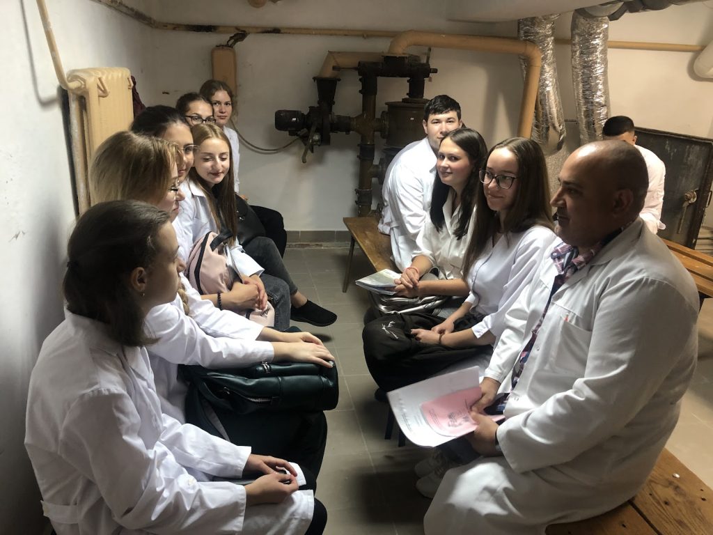
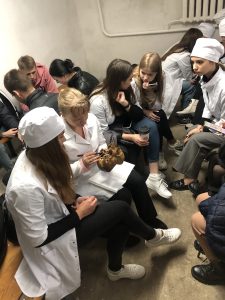
The educational process in the shelter of the Institute of Morphology
On April 17-19, 2019, the II stage of the All-Ukrainian Student Olympiad in the professionally oriented discipline “Human Anatomy” took place on the basis of the Educational and Scientific Institute of Morphology of the Ivan Horbachevsky Ternopil National Medical University.
Participants, organizers and jury members of the Olympiad
Performance of practical tasks by students participating in the Olympiad
Winners of the All-Ukrainian Student Olympiad in the professionally oriented discipline “Human Anatomy”
Photo in memory of Olympiad participants


